Gram staining is a widely used microbiological technique to differentiate bacterial species
into two main groups Gram-positive and Gram-negative, based on the structural differences
in their cell walls.
The technique, developed by Hans Christian Gram in 1884, involves a series of steps that
lead to the visualization of these differences under a microscope.
𝐏𝐫𝐢𝐧𝐜𝐢𝐩𝐥𝐞 𝐨𝐟 𝐆𝐫𝐚𝐦 𝐒𝐭𝐚𝐢𝐧𝐢𝐧𝐠
The principle of Gram staining is based on the differences in the thickness and composition
of the peptidoglycan layer in bacterial cell walls
𝐆𝐫𝐚𝐦 – 𝐏𝐨𝐬𝐢𝐭𝐢𝐯𝐞 𝐁𝐚𝐜𝐭𝐞𝐫𝐢𝐚
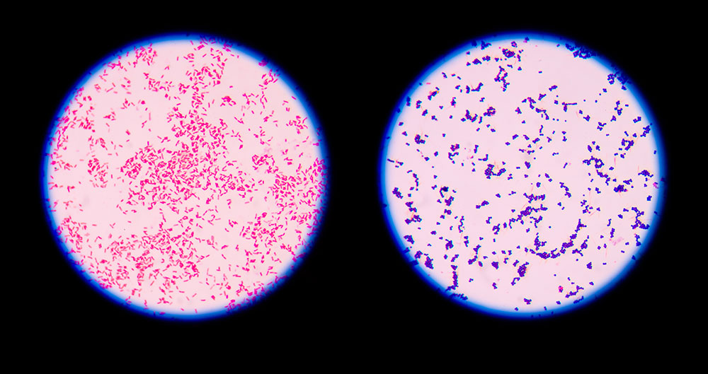
These bacteria have a thick peptidoglycan layer in their cell walls, which retains the primary
stain (crystal violet) after a series of treatments, appearing purple under the microscope.
𝐆𝐫𝐚𝐦 – 𝐍𝐞𝐠𝐚𝐭𝐢𝐯𝐞 𝐁𝐚𝐜𝐭𝐞𝐫𝐢𝐚
These bacteria have a thin peptidoglycan layer and an outer membrane that does not retain
the crystal violet stain after treatment with a decolorizer (alcohol or acetone). Instead, they
take up the counterstain (safranin) and appear red or pink.
𝐒𝘁𝗲𝗽𝘀 𝗶𝗻 𝗚𝗿𝗮𝗺 𝗦𝘁𝗮𝗶𝗻𝗶𝗻𝗴 𝗽𝗿𝗶𝗺𝗮𝗿𝘆 𝗦𝘁𝗮𝗶𝗻𝗶𝗻𝗴 (𝗖𝗿𝘆𝘀𝘁𝗮𝗹 𝗩𝗶𝗼𝗹𝗲𝘁)
A purple dye, crystal violet, is applied to the bacterial smear. This dye penetrates and stains
all the cells.
𝐌𝗼𝗿𝗱𝗮𝗻𝘁 (𝗜𝗼𝗱𝗶𝗻𝗲 𝗧𝗿𝗲𝗮𝘁𝗺𝗲𝗻𝘁)
Iodine is added as a mordant, which forms a complex with crystal violet, making the dye
molecules larger and more difficult to wash out of the cell.
𝐃𝗲𝗰𝗼𝗹𝗼𝗿𝗶𝘇𝗮𝘁𝗶𝗼𝗻 (𝗔𝗹𝗰𝗼𝗵𝗼𝗹 𝗼𝗿 𝗔𝗰𝗲𝘁𝗼𝗻𝗲 𝗧𝗿𝗲𝗮𝘁𝗺𝗲𝗻𝘁)
The smear is treated with alcohol or acetone, which dehydrates the thick peptidoglycan layer
in Gram-positive cells, trapping the crystal violet-iodine complex inside.
In Gram-negative cells, the alcohol disrupts the outer membrane and the thin peptidoglycan
layer, allowing the crystal violet-iodine complex to be washed out.
𝐂𝗼𝘂𝗻𝘁𝗲𝗿𝘀𝘁𝗮𝗶𝗻𝗶𝗻𝗴 (𝗦𝗮𝗳𝗿𝗮𝗻𝗶𝗻)
A red counterstain, safranin, is applied.
Gram-negative cells, which have lost the crystal violet, take up the safranin and appear red or
pink.
Gram-positive cells retain the purple color of crystal violet, so the safranin does not alter their
appearance.
𝐑𝐞𝐬𝐮𝐥𝐭𝐬
Gram-Positive Bacteria: Appear purple.
Gram-Negative Bacteria: Appear red or pink